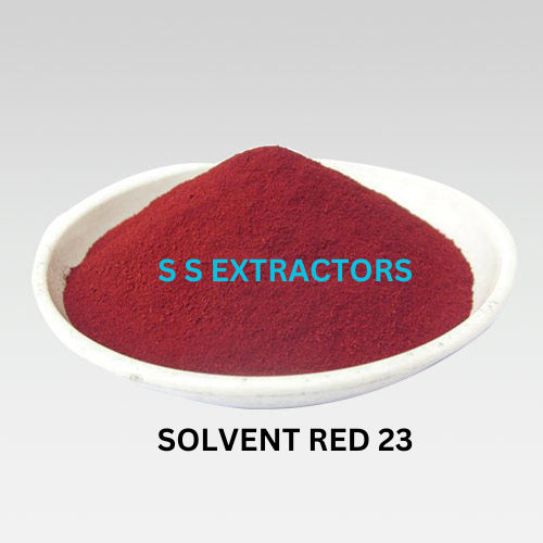
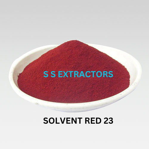
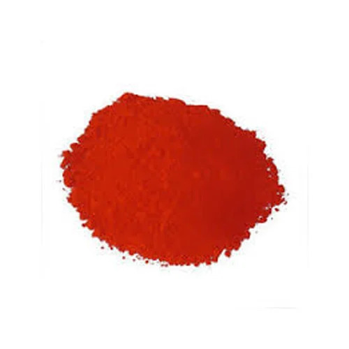
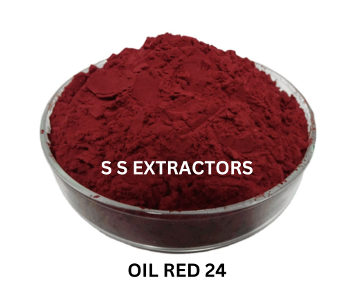
Oil Red 23 Dyes
Product Details:
- HS Code 85-86-9
- Ph Level 7 TO 8 (In Water)
- Solvent Color Red
- Density 0.12 TO 0.16 Kilogram per litre (kg/L)
- Solubility IN SOLVENT, OIL, WEX, VERY SOLUBLE IN BENZENE.
- Rubbing Resistance Dry
- CAS No 85-86-9
- Click to View more
Oil Red 23 Dyes Price And Quantity
- 10 Kilograms
- 400.00 INR/Kilograms
Oil Red 23 Dyes Product Specifications
- IN SOLVENT, OIL, WEX, VERY SOLUBLE IN BENZENE.
- Solvent Dye
- 85-86-9
- 0.12 TO 0.16 Kilogram per litre (kg/L)
- 85-86-9
- Dry
- Red
- Industrial
- Powder
- 7 TO 8 (In Water)
Oil Red 23 Dyes Trade Information
- 20000 Kilograms Per Month
- 3-7 Days
Product Description
Oil Red 23 is a synthetic dye that is commonly used in histology and biology for staining fat or lipids. It belongs to a group of dyes known as Sudan dyes, and specifically, it is part of the Sudan IV subgroup. These dyes are used to detect the presence of lipids, triglycerides, and neutral fats in tissues and cells.
Oil Red 23 is often used to stain frozen tissue sections, cell cultures, or other biological specimens to visualize and study the distribution of fat deposits. When applied, it selectively stains lipid-rich structures and appears as red or orange-red droplets or granules under a microscope.
Here's a basic overview of how Oil Red 23 staining works:
-
Fixation: The biological sample is typically fixed with a fixative like formaldehyde to preserve its structure.
-
Staining: The fixed sample is then incubated in an Oil Red 23 solution. The dye specifically binds to lipids, and lipid-rich structures within the sample will become stained.
-
Washing: After staining, the sample is washed to remove excess dye and non-specifically bound molecules.
-
Mounting: The stained sample can be mounted on a microscope slide and covered with a coverslip for examination under a microscope.
This staining technique is useful in various fields of research, including histology, pathology, and cell biology, to investigate fat distribution and lipid accumulation in tissues and cells. Researchers can use Oil Red 23 staining to identify and study conditions like lipid storage diseases, adipocyte differentiation, and fat content in various tissues.

Price:
- 50
- 100
- 200
- 250
- 500
- 1000+




 Send Inquiry
 Send SMS